Perio-Implant Advisory JOMI Clinical Pearls: Analyzing techniques for maxillary implant placement success
JOMIClinical Pearls is a regular column in Perio-Implant Advisorythat discusses articles from The InternationalJournal of Oral and Maxillofacial Implants (JOMI)—the official journal of the Academy of Osseointegration (AO)—as reviewed by a member of theAcademy’s Young Clinicians Committee (YCC). Quintessence Publishing publishes JOMI. Today’s studies are all pulled from Volume 30, Number 3, 2015.
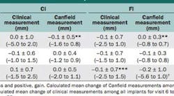
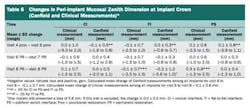
Today, I will tackle three different areas regarding the issues and information shared on maxillary implant placements. The first of these areas is the comparative trial of implants with different abutment interfaces to replace anterior, maxillary single teeth. The study includes 61 men and 80 women using implants that include 48 Conical Interface (CI), 49 Flat-to-flat Interface (FI), and 44 Platform Switch (PS). On all three types of implants, the preoperative study casts facilitated the surgical procedures, the augmentation procedures used protein 2 (rhBMP2), applied the flapless approach, and each implant manufacturer’s drilling protocols. Each implant was placed 3 mm apical of the planned gingival peri-implant mucosal zenith, and for provisionalization, the cases used a titanium abutment with provisional cement and without occlusal contacts. What the study shows us is that we achieve better results with immediate provisionalization when using the correct protocols. Also, CI had less marginal bone loss after one year compared with FI or PS. However, the changes in the buccal mucosal zenith position or papilla dimensions did not demonstrate big differences between the three implant designs (Tables 6 and 7).

