Saving teeth: They must not teach that as the first therapeutic response in dental school
As I look back on a consultation appointment I had with a patient named Martin five years ago, I realize how poignant that conversation has become in recent years. Martin presented to my office for consultation after seeing multiple dentists, including a few periodontists. He had been told by a general dentist he saw that he had a “severe amount of bone loss” and was in need of deep cleanings. In addition, “many of his teeth needed to be extracted,” since the bone loss was advanced and the “teeth were beyond repair.”
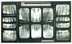
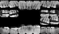
Figure 1: Full-mouth series taken in 2010, showing severe bone loss in the posterior maxillary molar and mandibular anterior incisor area.
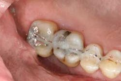
Figures 2a and 2b: Initial presentation in 2011 of the upper right and left molar area, showing advanced bone loss with furcation involvement on the molars.
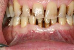
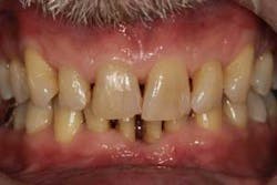
Figure 3: Initial presentation in 2011 of the mandibular anterior sextant, showing advanced hard- and soft-tissue loss.
Figure 4: Example of the type of splint used in the areas to be surgerized.
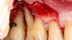
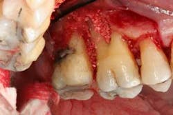
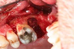
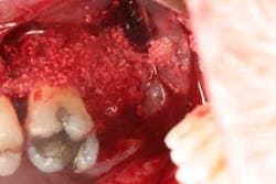
Figures 5a and 5b: Osseous surgery in the upper right quadrant with debridement of root surfaces and then regeneration.
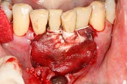
Figures 6a and 6b: Osseous surgery in the upper left quadrant with debridement of root surfaces, extraction of No. 15, and then regeneration.
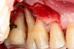
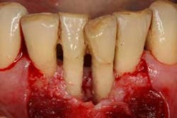
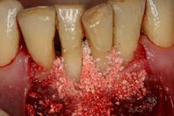
Figures 7a, 7b, and 7c: Osseous surgery in the mandibular anterior sextant with debridement of root surfaces and then regeneration.
Figure 8: Full-mouth series taken in 2016, showing radiographic evidence of bone regeneration in the areas surgerized.
Figure 9: Clinical presentation in 2016 of the mandibular anterior, showing increase in keratinized tissue and root coverage.
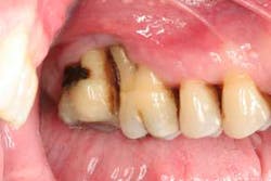
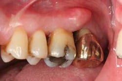
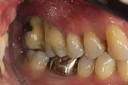
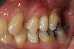
Figures 10 and 11: Clinical presentation in 2016 of the upper right and left molar area, showing an increase in tissue tone, soft- and hard-tissue levels, and a maintainable hygienic environment.
Martin was shocked by his diagnosis and dismayed by the hopeless prognosis he received. With a background in research as well as a questioning mind, Martin began to familiarize himself with periodontics and treatment options other than extraction and implants. He soon sought consultation with periodontal specialists to see if saving his teeth were an option. Although he was told that many of his teeth could be saved with periodontal treatment, he was again told that all of his maxillary molars needed to be extracted, including the four mandibular anterior teeth. In addition to extraction and site preservation, bilateral sinus augmentation with subsequent implant treatment would be necessary. Prosthetic restoration included multiple single-unit implant crowns, a four-unit implant bridge in the anterior, and various other cosmetic restorations. He was given a treatment plan that would cost close to six figures.
ADDITIONAL READING | Manage, repair, or regenerate periodontal disease?
When Martin came to my office (Disclaimer: 30%–40% of my practice involves preparing for and/or placing implants), he had a simple question: Can you save my teeth? When I told him that I thought it was possible as long as he was going to put as much effort into saving his teeth as I would need to, he thanked me and said, “I guess they don’t teach saving teeth as a first therapeutic response in dental school, since almost everyone I saw wanted to place implants.” Martin is better at describing his story in his own words, but the following is the clinical part of his journey through periodontal treatment.
At presentation, Martin was in his fifties with a medical history significant for controlled hypertension with no known food or drug allergies. He had been lackadaisical with his dental care in the past because of his busy schedule, but he was motivated to get his hygiene and treatment under control. He denied a history of smoking/alcohol/drug use. Based on his full-mouth series (figure 1) and clinical presentation, Martin had generalized, moderate bone loss with localized, severe bone loss in the posterior maxillary right and left quadrants (figures 2a and 2b) as well as the mandibular anterior incisor area (figure 3).
After explaining treatment options, a plan that was focused on saving as many teeth as possible was fabricated. This included four quadrants of scaling and root planing, splinting the areas of severe bone loss (figure 4), osseous surgery with hard- and soft-tissue regeneration, extracting tooth No. 15 (complete bone loss to the apical extent of the root), and diligent maintenance intervals.
ADDITIONAL READING | Understanding and managing peri-implant bone loss
Briefly, during osseous surgery, the upper right molars were treated with a combination of autograft and platelet-derived growth factor/Beta-tricalcium phosphate along with a porcine collagen graft (figures 5a and 5b). The upper left osseous surgery consisted of the same treatment (figures 6a and 6b), but also included the extraction of a hopeless tooth No. 15. Once again, the anterior osseous surgery consisted of the same regenerative materials as the former surgeries but with the addition of a porcine soft-tissue graft (figures 7a, 7b, and 7c). After the initial healing phase, the patient was placed on a strict home-care regimen and was seen every eight to 12 weeks for hygiene recare. The patient maintained excellent hygiene throughout the five-year follow-up period.
Today, five years after Martin was told that his maxillary molars and anterior incisors had to be removed, he has had a gain in bone levels (figure 8). He has had an increase in root coverage and keratinized tissue in the anterior incisal region (figure 9). In addition, he has no bleeding upon probing, probing depths of 2–3 mm (with the exception of a 4 mm probing distal to No. 3), and an overall gingival health that is far better than initial presentation (figures 10 and 11). After five years, I can now say to Martin, “Yes, it has been possible to save your teeth.”
Editor's note: Join us for the not-to-miss Perio-Implant Advisory Symposium on October 23, 2020. In this important virtual meeting, you’ll learn the newest skills it takes to save patients’ natural dentition. Bring your team and transform your practice. Conference schedule and special rates available at this link. Presented by Geistlich Biomaterials.
