Implant malposition: A new, easy-to-use radiographic aid for prevention
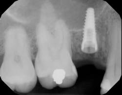
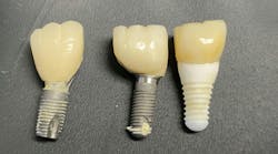
There are many reasons why dental implants ail or fail, with the most common complication arising from poor positioning of the dental implant fixture at the time of placement.1 Various surgical methods and techniques have been developed to try to correct implant malposition during surgery. In addition, there are many methods both restoring dentists and dental laboratories use to correct misplaced implants to give proper tooth anatomy and emergence profile.2
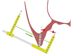
Although restorative and laboratory correction is possible, the best method continues to be prevention—i.e., placing the dental implant in the proper position at the time of surgery.3 Many clinicians still use freehand placement for single- and multiple-unit implant placement, and even with guided surgery, care must be taken when using information from perioperative radiographs to guide the surgical procedure. When taking guide pin radiographs during implant surgery, if the x-ray tube is not perpendicular to the radiographic film/sensor and the depth gauge, distortion of the image may occur (figure 1). This is especially important in a patient with limited opening and at the corners of the mouth in the canine area as there is a higher potential in these areas for alignment error.
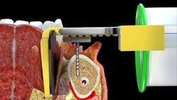
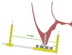
The Sniper X-Ray System is a simple device designed to ensure orthogonal x-ray beam alignment and correct osteotomy guide pin positioning so as not to provide false information to the implant surgeon during implant placement (figure 2).
By utilizing the Sniper X-Ray in combination with a conventional film/sensor holder, the implant surgeon can obtain consistent and accurate radiographs, resulting in greater measurement precision. This tool significantly improves the accuracy of intraoperative radiographs, allowing for more precise reevaluation during the dental implant surgical procedure (figure 3).
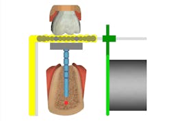
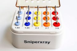
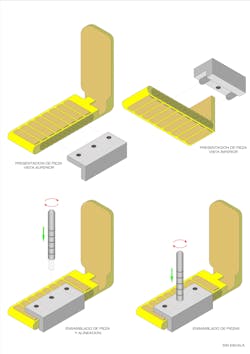
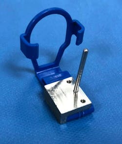
The guide pins come in a variety of osteotomy sizes, allowing the user to take serial radiograph films during the procedure if needed (figure 4). This system consists of a base that attaches to the radiographic film holder. The base can be fixed or inserted on both sides of the film holder, allowing for a secure and stable attachment of the Sniper X-Ray tool (figures 5 and 5a). Assembly and disassembly are easy, and the system can be sterilized.
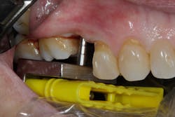
Appropriate alignment of the x-ray beam is a critical factor in obtaining clear and accurate radiographic images, reducing distortion, and improving diagnostic quality during dental implant osteotomy preparation. When diagnostic radiographic data transfer is accurate, the dental implant has a higher probability of being placed appropriately (figures 6 and 6a).
Watch this video to see how this system is used during dental implant surgery.
Disclosure: Dr. Arguelles is the inventor and owner of the Sniper X-Ray System.
Editor’s note: This article originally appeared in Perio-Implant Advisory, a chairside resource for dentists and hygienists that focuses on periodontal- and implant-related issues. Read more articles and subscribe to the newsletter.
References
- Monje A, Galindo-Moreno P, Tözüm TF, del Amo FSL, Wang HL. Into the paradigm of local factors as contributors for peri-Implant disease: short communication. Int J Oral Maxillofac Implants. 2016;31(2):288-292. doi:10.11607/jomi.4265
- Chatterjee A, Ragher M, Patil S, Chatterjee D, Dandekeri S, Prabhu V. Prosthetic management of malpositioned implant using custom cast abutment. J Pharm Bioallied Sci. 2015;7(Suppl 2):S740-S745. doi:10.4103/0975-7406.163528
- Arisan V, Karabuda ZC, Ozdemir T. Accuracy of two stereolithographic guide systems for computer-aided implant placement: a computed tomography-based clinical comparative study. J Periodontol. 2010;81(1):43-51. doi:10.1902/jop.2009.090348
Angel Eyaralar Arguelles, MD, DDS, received his medical degree from the Universidad de Oviedo. He received his doctor of dental surgery degree from the Iberoamericana University. He is a specialist in plastic and reconstructive surgery training at the José Guerrerosantos Reconstructive Surgery Institute. He is further trained in dental implant surgery from the Brånemark Osseointegration Centre in Madrid, Spain. He has a private practice exclusively dedicated to dental implant surgery in Asturias, Spain.
Scott Froum, DDS, a graduate of the State University of New York, Stony Brook School of Dental Medicine, is a periodontist in private practice at 1110 2nd Avenue, Suite 305, New York City, New York. He is the editorial director of Perio-Implant Advisory and serves on the editorial advisory board of Dental Economics. Dr. Froum, a diplomate of both the American Academy of Periodontology and the American Academy of Osseointegration, is a volunteer professor in the postgraduate periodontal program at SUNY Stony Brook School of Dental Medicine. He is a PhD candidate in the field of functional and integrative nutrition. Contact him through his website at drscottfroum.com or (212) 751-8530.
About the Author

Angel Eyaralar Arguelles, MD, DDS
Angel Eyaralar Arguelles, MD, DDS, received his medical degree from the Universidad de Oviedo. He received his doctor of dental surgery degree from the Iberoamericana University. He is a specialist in plastic and reconstructive surgery training at the José Guerrerosantos Reconstructive Surgery Institute. He is further trained in dental implant surgery from the Brånemark Osseointegration Centre in Madrid, Spain. He has a private practice exclusively dedicated to dental implant surgery in Asturias, Spain.
