Guided bone regeneration treats dental implant lesions
Oral implant surgery is complex and not without complications, one of which is an implant periapical lesion (IPL). If the lesion site becomes infected, it can lead to an abnormal growth, persistent inflammation and tenderness. However, a procedure that allows complete bone regeneration at the implant related lesion site shows promise in treating the resulting bone defect and infection.ADDITIONAL READING |Study finds that shorter waiting time between dental procedures is adequate IPL develops rapidly after implant surgery and is treated with a second reparative surgery, in combination with antibiotic use. Surgeons have tried various treatments, such as using hand tools to enucleate the lesion, placement of bovine bone mineral to replace the diseased bone, and using an enamel mixture to help strengthen the surrounding tissues. Results from these treatments have been mixed. Some treatments have been successful, while others resulted in the lesion progressing, and in others the implant was lost.ADDITIONAL READING |Atrophic patients have more options with new dental implant In a Journal of Oral Implantologycase study titled “Active implant periapical lesion: a case report treated via guided bone regeneration with a five-year clinical and radiographic follow-up,” surgeons reported on using guided bone regeneration (GBR) principles to completely remove the lesion and any subsequent infections.
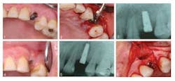
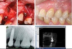
A 43-year-old female presenting with a symptomatic left, first premolar was a candidate for dental implant treatment and scheduled for an immediate implant placement following tooth extraction. After surgery she was prescribed antibiotics. She was seen three months later due to pain at the implant site, which revealed a sinus tract related to the implant. Additionally, there was a “soft spot” due to edema and bone loss. She was prescribed another course of antibiotics and returned in four days. At that time, a tetracycline paste was created and placed on the defect and around the implant for three minutes, then removed. In two months, a transitional crown was placed, with placement of the final six months later. At the subsequent one-, two-, six-month, one- and five-year appointments, no pain was reported and complete bone fill (see photo below) in to the previous lesion area was stable.
IPL is a rare disorder, affecting approximately 0.26% of the population receiving implants. There are varying reasons for its cause, and it can sometimes be misdiagnosed or confused with retrograde peri-implantitis. The combination of antibiotics and GBR principals has shown to be an effective way of treating IPL, keeping the implant intact, and creating a complete bone fill at the lesion site. This case study appears to be the first of its kind, so further research will be needed to confirm findings.ADDITIONAL READING |Less invasive approach to dental implants allows heart patients to continue anticoagulation therapy Full text of the article, “Active implant periapical lesion: a case report treated via guided bone regeneration with a five-year clinical and radiographic follow-up,” Journal of Oral Implantology, 2014;40(3) is available here. For more information about the Journal of Oral Implantology, vist their website.


