Strategic retention of an ailing premolar with severe periodontitis using regenerative therapy: a case report
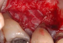
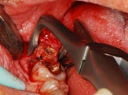
Fig. 2 — This view is taken after root planing. The lesion is a combined 1-2 wall that extends from the distal to the palate. What complicated care was that the tooth could not be extracted without incurring considerable costs. If the patient wished to replace this tooth with a fixed partial denture, she would need to have the existing seven-unit splint anterior to this tooth removed. If an implant was the desired option, the patient would need to undergo elevation of the sinus and — at the very least — wear a removable appliance until an implant could be placed and integrated, something she wished to avoid. The decision was to perform occlusal equilibration to eliminate fremitus in centric occlusion and excursive movements. Regenerative surgery was scheduled for this area that included elevation of full thickness flaps, meticulous debridement of the roots with hand and ultrasonic instruments, and root modification with tetracycline solution (Fig. 2). The root was then treated with a solution of a highly purified protein, recombinant platelet-derived growth factor (rh-PDGF-BB). The remaining/containing walls of the intrabony lesion were not ideal for space maintenance to stabilize a clot as their morphology had one to two wall components. To regenerate this site, an allograft of mineralized, freeze-dried bone (FDBA) (LifeNet Health, Virginia Beach, Va.) hydrated by rh-PDGF-BB was carefully placed into the defect to act as a biologic scaffold/clot stabilizer (Fig. 3). To further contain this graft-biologic composite, a barrier made from human chorion-amnion (BioXclude, Snoasis Medical Inc., Denver, Colo.) was placed over the graft-biologic and the flaps (Fig. 4) were secured with 6-0 expanded polytetrafluoroethylene (ePTFE) (W.L. Gore & Associates, Flagstaff, Ariz.), assuring complete closure of the site (Figs. 5 and 6).
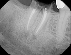
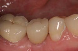
Postoperative infection control consisted of a systemic antibiotic — amoxicillin taken for the first seven days and the topical use of chlorhexidine mouth rinse applied to the site for the first month. For analgesia and the reduction of inflammation, ibuprofen 600 mg was prescribed for the first four to five days. The patient was seen approximately every 14 days for postoperative treatment during the three months. Sutures were removed at the first visit. She was then seen monthly up to the sixth postsurgical month. Postoperative visits included plaque debridement, polishing to remove stains, and oral hygiene reinforcement. The early frequency of visits was imperative as the patient had to refrain from both brushing and flossing of the treated area to provide wound quiescence until the clot had stabilized. After the first 30 days, mechanical oral hygiene was reinstated and the patient resumed her mechanical plaque control along with rinsing with an essential oil mouth rinse twice daily. Endodontics was completed at 28 days postsurgery to treat the apical lesion, and the patient resumed her typical three-month interval of maintenance care after the initial six months of stringent postoperative visits. The bone loss at the distal of this tooth has been substantially recaptured and the attachment level and probing depths have been restored to maintainable levels of 3 mm to 4 mm. Two years later, the bone levels have remained stable and the prognosis for this tooth has considerably improved (Figs. 7 and 8).

