Dental implants: The importance of patient compliance with immediate loading
Immediate placement and immediate loading have been buzzwords in implant dentistry for decades. Patient demand for immediate gratification has increased in the age of the I-generation. Implant dentistry has not been immune to the ‘I want it now’ and has since responded with immediate placement after extraction followed by immediate loading of the prosthesis. Immediate placement and load has enjoyed similar success rates as compared with conventional delayed placement; however, this technique for dental implants requires higher patient compliance. (1) Smoking, parafunction, hygiene, and overloading are all patient factors (2) that are hard to control and can lead to higher failure rates when dealing with immediate-load situations. Overload is certainly one factor that needs to be reinforced with patients when loading an implant immediately after placement. (3) We assume that patients will be compliant because they want treatment to work; however, that is not always the case. The following case example demonstrates a scenario that is possible when a patient is not compliant after an immediate-loading protocol.
ALSO BY DR. PETER MANN |Narrow diameter dental implants in the esthetic zone
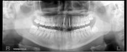
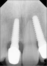
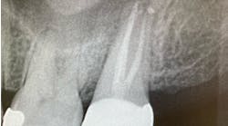
Fig. 1: Preop PAN, June 22, 2013.One-piece implants No. 8 and No. 10 were placed in a flapless manner. Excellent initial stability was achieved (Fig. 2). Patient was temporized out of occlusion (Fig. 3). Postop instructions were once again reviewed and antibiotics were prescribed.
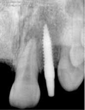
Fig. 2: Implant placement — 3.0 x 14 mm implant placed on No. 10.
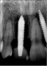
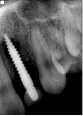
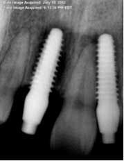
Fig. 3: Implant temporization No. 8 and No. 10 — 3.7 mm x 13 mm implant placed on No. 8.Patient returned one month later after returning from a cruise, complaining that her “implants felt loose.” She was very honest and admitted to not taking the antibiotics and not following any dietary restrictions while on vacation, because “they felt so strong.” Periapicals taken that day showed a catastrophic failure, with large areas of bone loss surrounding both implants (Fig. 4).
After explaining to the patient that because of her noncompliance the implants would have to be removed, she decided to wait two weeks for the procedure. She was then anesthetized and the implants removed. Fortunately, this patient had a sufficient amount of bone to allow wider implants to be placed. Upon careful examination and probing, there were no perforations found in the bone.
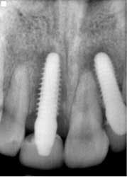
Implant sites were curetted well, then disinfected with Chloramine-T saturated gauze, followed by irrigation with 2% chlorhexidine and water. Bone grafting was not deemed necessary. Both implants No. 8 and No. 10 were replaced with TRX-OP 4.5 x 13 mm (Fig. 5). The patient was informed that because implants placed into previously failed sites have a lower success rate, absolute adherence to postoperative instructions was critical to any chance of success (4). Instructions were once again given to follow dietary restrictions and take the antibiotics as prescribed. The patient was seen six weeks postoperatively (Fig. 6) and set to have the final prosthesis inserted six months post-reimplantation.Fig. 5: No. 8 and No. 10 — implants replaced by 4.5 x 13 mm and retemporized.
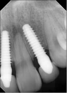
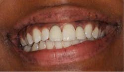
Fig. 6: Postoperative six weeks later.The patient returned for a follow-up eight months later (Fig. 7) and claimed she was careful not to bite into anything with her front teeth and wanted to know if they could be restored with permanent crowns. Impressions were taken and sent to the lab for fabrication of implant crowns. One year post-insertion, the final crowns and radiographs were taken (Figs. 8 and 8a).
Fig. 7: Healing eight months post-reinsertion.
Fig. 8: One year post-insertion — final crowns.
Fig. 8a: One year post-insertion — final radiographs.
References
1. Luongo et al. Immediate functional loading of single implants: a one-year interim report of a five-year prospective multicentre study. Eur J Oral Implant 2014 Summer;7(2):187-199.
2. PEsquivel-Upshaw J et al. Peri-implant complications for posterior endosteal implants. Clin Oral Implants Res. Sept. 27, 2014.
3. Duyck J et al. The effect of loading on peri-implant bone: a critical review of the literature.J Oral Rehabil. Oct. 2014;41(10):783-794. doi: 10.1111/joor.12195. Epub 2014 Jun 3.
4. Wang F et al. Intermediate long-term clinical performance of dental implants placed in sites with a previous early implant failure: a retrospective analysis. Clin Oral Implants Res. Nov. 13, 2014.


