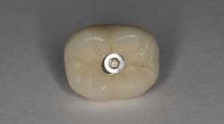
Prosthetic screw loosening: An innovative technique to manage a common complication with dental implants
Introduction
Recent systematic reviews have shown that the success rate of dental implant-supported crowns does not appear to be affected by retention type. (1) Nevertheless, screw-retained and cement-retained restorations offer relative advantages and disadvantages with respect to biological and technical complications. (2) Retention type should be chosen based upon an understanding of these relative advantages and disadvantages in order to minimize the potential for future implant complications. (3)
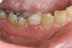
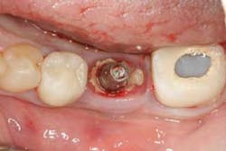
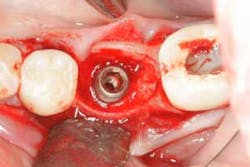
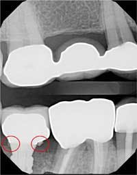
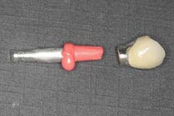
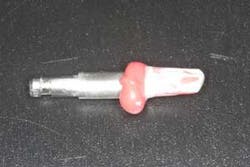
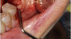
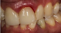
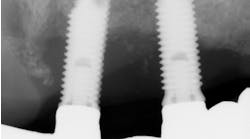
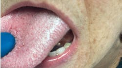
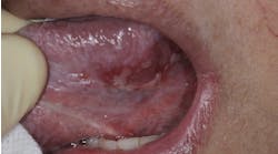
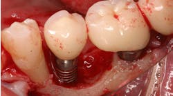
Studies have shown that cement-retained crowns have a higher incidence of peri-implant mucositis (inflammation of the peri-implant soft tissue: Figure 1) and peri-implantitis (inflammation of the peri-implant soft and hard tissue: Figures 2 and 3), likely due to the presence of excess cement following crown cementation. (4) All measures should be taken to avoid the extrusion of excess cement when cementing implant crowns. The use of a radiopaque cement is recommended to detect any excess cement radiographically immediately following cementation (Figure 4). (5) A custom cement analog also can be used to help minimize the chances of excess cement extrusion (Figures 5 and 6). (6)
Figure 1:Inflammation of the buccal soft tissue with suppuration emerging from the sulcus.
Figure 2:Abundance of excess cement noted in the peri-implant sulcus once crown was removed.
Figure 3:Note the extent of bone loss around the dental implant induced by the presence of excess cement.
Figure 4:Excess cement detected radiographically postcementation.
Figure 5:An abutment replica was fabricated prior to cementation.
Figure 6:After loading the crown with cement, the crown is seated on the abutment replica to exude excess cement and evenly distribute cement within the crown.
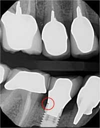
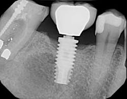
Technical complications such as screw loosening and lack of passive seating of the framework (Figure 7), however, are more common in screw-retained restorations. (3) Nevertheless, screw-retained crowns offer a major advantage over cement-retained restorations because they are retrievable. Efforts have been made to increase the retrievability of cement-retained restorations by using temporary cements instead of permanent cements. Despite this, the retrievability of cement-retained crowns is not predictable. (7)
Figure 7:Note the lack of complete seating of the restoration detected radiographically.
ADDITIONAL READING |Etiology, prevention, and treatment of screw loosening and fracture
When abutment screw loosening occurs with a cement-retained crown, the clinician is faced with a difficult challenge. As opposed to a screw-retained restoration, which offers easy access to the abutment screw for tightening, a cement-retained crown is typically still cemented to the loose abutment and cannot be removed intraorally without cutting the crown. If the crown-abutment unit can be removed from the implant in one piece, the crown may be able to be “uncemented” from the abutment by placing it in a porcelain oven. This requires the dentist to send the crown to the lab, leaving the patient without a crown for several days until it comes back from the lab with the abutment. The following case report illustrates a technique to reseat a cement-retained crown-abutment unit that loosened from the implant by converting a cement-retained crown into a screw-retained crown at the very same appointment.
Case report
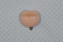
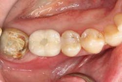
A 65-year-old female presented with a cement-retained implant crown No. 30 on an external hex implant, which became loose (figure 8). The crown had been in function for 15 years. Fortunately, the cement-retained crown was able to be removed, but it was still attached to its abutment and abutment screw (figure 9) and, therefore, was not able to be recemented.
Figure 8:Loose implant crown.
Figure 9:Cement-retained crown removed still attached to its abutment.
There were three options available. One option was to take a new impression and fabricate a new abutment and crown. The obvious disadvantage of this option is the added cost to the patient and the fact that the patient would have to be without a crown for several days.
A second option was to send the crown-abutment unit to the laboratory and have it placed into a porcelain oven to burn out the cement. The crown could then be separated from the abutment so that the abutment could be reseated and the crown recemented. This is not always possible, however, since there is a risk that the porcelain may distort when it is placed back in the oven. Additionally, this option also requires that the patient be without a crown for several days.
In this case, to minimize the cost to the patient and avoid the need to be without a crown for several days, the dentist decided to convert the cement-retained crown into a “screw-retained” restoration.
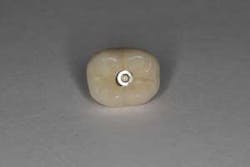
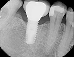
The screw channel of the abutment was accessed by carefully drilling a hole in the center of the occlusal surface using a cross-cut 557 bur (Brasseler) and progressively widening it until the screw channel was accessed (figure 10). Once the screw channel was accessed, the cavit and cotton blocking the screw were removed and the screw was visualized. A driver was placed into the channel to engage the screw and the cement-retained crown was seated on the implant and “screwed” back in place. Complete seating of the restoration was verified radiographically (figure 11). The screw was torqued to 25 Ncm and the access channel was filled with Teflon tape and closed with composite (figure 12). The patient was able to leave with the crown securely in place.
Figure 10:Screw channel accessed.
Figure 11:Complete seating of the restoration confirmed radiographically.
Figure 12:Composite sealing new access hole.
Conclusion
Both screw-retained and cement-retained implant-supported crowns can be used successfully to restore implants. The clinician should be aware of the relative advantages and disadvantages of each option with respect to biological and technical complications, so that every measure can be taken to avoid future complications and manage these complications when they occur. If a screw-retained option is selected, complete seating of the restoration must be confirmed at the time the restoration is placed. If a cement-retained option is selected, efforts must be taken to avoid extrusion of excess cement at the time of cementation to prevent cement-induced peri-implant disease. The above case report illustrates the relative disadvantage of a cement-retained option with respect to retrievability and presents an innovative technique to reseat the restoration with minimal cost and time to the patient.
References
1. Wittneben JG, Millen C, Brägger U. Clinical performance of screw- versus cement-retained fixed implant-supported reconstructions—a systematic review. Int J Oral Maxillofac Implants. 2014;29(suppl):84–98. doi: 10.11607/jomi.2014suppl.g2.1.
2. Millen C, Brägger U, Wittneben JG. Influence of prosthesis type and retention mechanism on complications with fixed implant-supported prostheses: a systematic review applying multivariate analyses. Int J Oral Maxillofac Implants. 2015;30(1):110–124. doi: 10.11607/jomi.3607.
3. Shadid R, Sadaqa N. A comparison between screw- and cement-retained implant prostheses. A literature review. J Oral Implantol. 2012;38(3):298–307. doi: 10.1563/AAID-JOI-D-10-00146.
4. Wilson, TG Jr. The positive relationship between excess cement and peri-implant disease: a prospective clinical endoscopic study. J Periodontol. 2009;80(9):1388–1392. doi: 10.1902/jop.2009.090115.
5. Pette GA, Ganeles J, Norkin FJ. Radiographic appearance of commonly used cements in implant dentistry. Int J Periodontics Restorative Dent. 2013;33(1):61–68.
6. Frisch E, Ratka-Krüger P, Weigl P, Woelber J. Minimizing excess cement in implant-supported fixed restorations using an extraoral replica technique: a prospective 1-year study. Int J Oral Maxillofac Implants. 2015;30(6):1355–1361. doi: 10.11607/jomi.3967.
7. Mehl C, Harder S, Shahriari A, Steiner M, Kern M. Influence of abutment height and thermocycling on retrievability of cemented implant-supported crowns. Int J Oral Maxillofac Implants. 2012;27(5):1106–1115.
