Literature review: Peri-implant disease
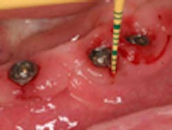
Peri-implant disease can be divided into peri-implant mucositis and peri-implantitis. Peri-implant mucositis is noted by the presence of bleeding, pain, or suppuration. Peri-implantitis may present the same way but is marked by the observation of radiographic bone loss beyond anticipated levels. The prevalence of peri-implant disease has been cited as high as 80% for peri-implant mucositis and between 28% and 56% for peri-implantitis (Lindhe et al, Ziztmann et al). It is caused by the bacteria in plaque and calculus similar to periodontal diseases around natural teeth (Luterbacher et al). Although the disease is caused by bacteria, there are other important risk factors for peri-implant disease, most notably tobacco use (Karbach et al). One way to reduce the incidence of the disease is to have a minimum of 2 mm of keratinized tissue around the implant to reduce plaque accumulation (Schrott et al). Once peri-implant mucositis has developed, there are nonsurgical treatment methods available, including mechanical debridement, antimicrobial rinse, and local or systemic antibiotics (Renvert et al). There is no consensus in the literature about the surgical treatment of peri-implantitis; however, some studies are showing promise (Froum et al).
RELATED |A new tool in the fight against peri-implant disease
RELATED |Literature review: Emergence profiles for dental implants
