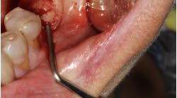
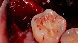
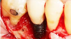
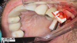
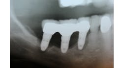
Sinus means “pocket” in Latin. Humans have these air-filled spaces that surround the nasal cavity called paranasal sinuses. The four-paired paranasal sinuses include frontal and ethmoid sinuses between the eyes, sphenoid sinuses behind the ethmoid, and maxillary sinuses surrounding the nasal cavity. The maxillary sinuses are the largest of the paranasal sinuses. Although not proven, the biological roles of sinuses include decreasing the relative weight of skull, increasing voice resonance, providing a buffer against blows to face, insulating structures, and humidifying/heating inhaled air. (1)
The maxillary sinus is pyramidal in shape. The base of the pyramid is the medial wall of the sinus that is also the lateral wall of the nasal cavity, and its apex is pointed toward the zygomatic bone. The roof of the sinus is also the floor of the orbit. The average volume of a sinus is about 15 ml (range between 4.5 and 35.2 ml). The maxillary sinus maintains its overall size while the posterior teeth remain in function as the size expands with age, especially when posterior teeth are lost. This process is called pneumatization and is possibly the result of atrophy caused by reduced strain from occlusal function.
The membrane that lines the walls of the maxillary sinus is called Schneiderian membrane. It is multilayered and 0.13 mm to 0.5 mm in thickness. The layers include the respiratory epithelium (pseudo-stratified ciliated columnar epithelium) that covers a loose, highly vascular connective tissue and periosteum. The healthy maxillary sinus is self-maintaining by postural drainage and actions of the ciliated epithelial lining, which propels bacteria toward the ostium. The ostium is a nonphysiologic drainage port high on the medial wall that opens into the nasal cavity between the middle and lower nasal conchae (hiatus semilunaris).
At its highest point, the ostium is located 30 mm above the floor and serves as an anatomic rationale for the sinus floor elevation procedure. Sinus elevation may improve the symptoms of sinusitis and congestion by bringing the floor of sinus closer to the drainage port, and the grafting procedure does not interfere with normal sinus function. (1)
The blood supply to the maxillary sinus is derived primarily from the maxillary artery. The posterior superior alveolar and infraorbital arteries anastomose in the bony lateral wall. (2) On average, the intraosseous anastomose (IA) occurs in 100% of the cases and 19 mm from the alveolar bone crest, whereas the extraosseous anastomose (EA) occurs in about 40% of the cases and 23 mm from the alveolar bone crest. There may be one or more septa dividing the maxillary sinuses, called Underwood’s septa. (3,4,5) It occurs about 30% of the time and in the anterior region (most commonly between the second premolar and first molar) about 77% of the time. The mean height of the septae is 7.9 mm (range 0 to 17 mm). (3,4,5)
As mentioned above, the maxillary sinus provides a challenge in implant dentistry because of the reduced bone volume, which is due to alveolar bone resorption and pneumatization of the sinus cavity. Several ways to avoid the sinus cavity is to use a short implant, tilt the implant mesially or distally, use a long zygomatic implant, and/or shorten the dental arch with premolar occlusion. With the premolar occlusion, 50% to 80% of chewing capacity is maintained. (6) However, over the years, techniques have been developed to augment the sinus floor.
Philip Boyne is the first to report the elevation of the maxillary sinus floor for preprosthetic reasons. The maxillary sinus was augmented prior to a tuberosity reduction to increase the interarch distance and create a more symmetric maxillary arch for denture prosthesis. (7) Boyne also reported on the two-stage elevation of the maxillary sinus floor as a preparation for the placement of blade implants. He grafted the maxillary sinus with autogenous particulate iliac bone, and then placed blade implants three months later. (8)
Hilt Tatum suggested the crestal approach for sinus floor elevation with subsequent implant placement. A “socket former” was used to prepare the implant site and create green-stick fracture of the sinus floor. A root-formed implant was placed and allowed to heal in a submerged way. (9)
Robert Summers described another crestal approach — BAOSFE (Bone-Added Osteotome Sinus Floor Elevation) — using tapered osteotomes with increasing diameters. Adjacent bone was compressed by pushing and tapping as the sinus membrane was elevated. Autogeneous, allogenic, or xenogenic bone grafts were added to increase the volume below the elevated sinus membrane. Using this approach, Summers reported a 96% success rate at 18 months after loading 173 press-fit submerged implants. (10)
There are two major approaches to elevate the sinus floor: lateral window and transalveolar approach. The lateral window approach can be one- or two-stage techniques for implant placement; the transalveolar approach is a one-stage technique, mainly based on available residual bone and the possibility of achieving the primary stability of the implant.
Author bio
Dr. Joo H. Kim received his DDS from New York University College of Dentistry, where he was elected to membership in Omicron Kappa Upsilon (OKU), the National Dental Honor Society. Dr. Kim is currently a chief postgraduate periodontology resident at the SUNY Stony Brook School of Dental Medicine.
References
1. Lindhe J, Lang N, Karring T. Ch. 50: Elevation of the maxillary sinus floor. Clinical Periodontology and Implant Dentistry, Fifth Edition 2008.
2. Solar P, Geyerhofer U, Traxler H, Windisch A, Ulm C, Watzek G. (1999). Blood supply to the maxillary sinus relevant to sinus floor elevation procedures. Clinical Oral Implants Research 1999; 10(1), 34-44.
3. Underwood AS. An inquiry into the anatomy and pathology of the maxillary sinus. Journal of Anatomy and Physiology1910; 44:354-369.
4. Ulm CW, Solar P, Gsellmann B, Matejka M, Watzek G. The edentulous maxillary alveolar process in the region of the maxillary sinus — a study of physical dimension. Journal of Oral and Maxillofacial Surgery 1995; 24:279-282.
5. Kim MJ, Jung UW, Kim CS, Kim KD, Choi SH, Kim CK, Cho KS. Maxillary sinus septa: prevalence, height, location, and morphology. A reformatted computed tomography scan analysis. Journal of Periodontology 2006; 77(5):903-908.
6. Kayser AF. Shortened dental arches and oral function. Journal of Oral Rehabilitation 1981; 8(5):457-462.
7. Boyne PJ. Restoration of osseous defects maxillofacial causalities. Journal of American Dental Association 1969; 78:767-776.
8. Boyne PJ, James R. Grafting of the maxillary sinus floor with autogeneous marrow and bone. Journal of Oral Surgery 1980; 38:613-618.
9. Tatum H. Maxillary and sinus implant reconstructions. Dental Clinics of North America 1986; 30(2):207-229.
10. Summers RB. (1994). A new concept in maxillary implant surgery: the osteotome technique. The Compendium of Continuing Education in Dentistry 1994; 15(2):152-162.

