How a perfect, implant-retained anterior bridge can cause facial pain
Here is a problem that you might encounter. Reading this will help you and your patient through a potentially difficult situation.
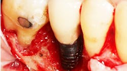
A patient has two implants placed in the No. 7 and No. 10 positions. Surgery goes fine. Tissues are healthy. Implants are perfectly placed as verified by computed tomography (CT). Uncovering is uneventful with plenty of keratinized tissue on the facial, and there is a great emergence profile.
The patient returns to her restorative dentist for fabrication of the final abutments and a temporary, fixed prosthesis. Custom abutments are perfect. They are zirconia that are shaded perfectly so that the margins can be slightly supragingival, keeping esthetics at their optimum without risking subgingival cement.
Abutments go in fine without the need for anesthetic and are torqued at 30 Ncm with no discomfort to the patient. The dentist cements in a temporary, fixed bridge to test for function and esthetics. There is minimal cement and slight blanching of the tissue in the pontic area to develop some tissue remodeling.
Do you get the picture? Everything up to this point is perfect.
Here's where the changes occur: The patient returns to the office with a complaint of pain in the implants. She has headaches, and it feels as if her teeth are being squeezed together. The dentist removes the temporary, fixed bridge and the pain almost completely goes away. So the dentist, being conscientious and assuming that the problem is the tissue pain caused by the pontics, relieves the pontic area completely and recements the bridge. The patient is fine.
ADDITIONAL READING |Diagnosing between ailing and failing implants: The haste to remove salvageable implants
She returns the next day with the same complaint, however. It feels as if her teeth are being squeezed together, and it's giving her a bigger headache. The general dentist says, "Maybe the acrylic shrunk a bit. I'll relieve the internal aspect of the bridge.” He removes it.
The patient returns the next day with the same complaint. The dentist removes the abutments, puts in some short, healing abutments, and modifies the temporary partial that the patient wore during the healing period. Symptoms are much better with just a little of the discomfort remaining, but she can live with it. What's the diagnosis? More importantly, what do you do next?
The answer to this question may fall into an area that we were never taught about in dental school and one that isn't often recognized in traditional dentistry or medicine. The answer may be an interference with the cranial rhythm.
What is a cranial rhythm? The cranial rhythm is identified in two disciplines: cranial osteopathy and chiropractic. Each of the cranial bones of the skull articulate with each other at the cranial sutures. And contrary to what we may have been taught, the sutures do not completely fuse in adulthood. Remember when you studied the cranial bones? They could be studied as individual bones, complete with jagged and irregular suture lines.
So if the cranial bones are not fused, there must be a purpose for their independence. That purpose is all tied in to a concept called "cranial rhythm" and the "cranio-sacral pump." As you know, our central nervous system is bathed in cerebrospinal fluid. But the cerebrospinal fluid just doesn't sit there. It needs a pumping mechanism to keep it flowing. That pumping mechanism is created by the rhythmic contraction of the cranial bones at the top and the sacrum at the bottom. The rhythmic motion of these two terminals is called the cranio-sacral pump. The rhythm is estimated to be about four cycles per minute.
Now here's the problem. When we have a fixed dental restoration that crosses the midline, the restoration has the potential to impede the flexion of the mid-palatal suture, which, in turn, impedes the flexion of the other cranial bones, disturbing the cranial rhythm. This can be reflected as headaches and as a squeezing feeling in the upper anterior sextant. It can happen not only with implants, but also with natural teeth. Most patients may never feel this, but for some, it can be a nightmare. And, of course, it is even worse when the patient is told that it's all in his or her head. In fact, it is in their head as a iatrogenic impedance of the cranial rhythm.
What do you do when you see this?
- Believe your patient. Don't write symptoms off as psychosomatic or worse.
- Remove the fixed bridge. This is a reason to use temporary cement rather than permanent cement.
- If the patient gets relief with the bridge off, refer to a cranial chiropractor or cranial osteopath in your community. Keep the patient in a removable prosthesis during this time.
- Once the patient has achieved complete relief, the prosthesis may then need to be revised so that it doesn't cross the midline. A semiprecision attachment that allows for mid-palatal suture movement may be an answer. Another answer may be a new prosthesis with more abutments so that the two prostheses don’t have to cross the midline.
It is the patient’s viewpoint that is paramount. The patient will tell you the answer. All we have to do is observe, ask, and listen.


