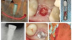
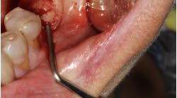
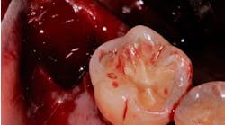
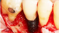
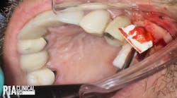
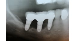
The hole left by a pulled tooth is more than just a place where food can get caught and the tongue can “worry” the gap. It is also a place where disease can weaken bone. Surgeons typically fill this hole with socket grafting material, but a barrier placed over the graft may help the bone regrow even faster.
ADDITIONAL READING |Does your dental extraction socket need a bone graft: A decision matrix
A study in the current issue of the Journal of Oral Implantology looks at a new type of barrier membrane, known as porcine collagen that has been recently introduced in the United States. The intent was to find out how quickly a bone graft can develop when porcine collagen is placed over the grafted tooth socket.
Read the abstract.
Socket grafting is one of the most frequently performed procedures in oral surgery. After a tooth is pulled, the tooth socket in the jawbone where the tooth had been anchored can rapidly shrink and make it impossible to place a dental implant. To prevent this, the surgeon fills the hole with a bone grafting material that combines with the natural bone to rebuild or preserve the bone’s strength. A barrier membrane, such as porcine collagen, can keep the gum from growing into the space.
The current study involved 14 patients who needed to have one or more teeth replaced. Once the teeth were removed, the sockets were filled with particulate allograft bone and covered with a layer of porcine collagen. After 16 weeks, the sites were examined and dental implants were placed. The results showed a wide range of new bone growth in the treated sockets, from 1.8% to 43%. The new bone formation averaged 11.2% among the study group. At the same time, the barrier of porcine collagen helped to prevent soft tissue from growing into the space. It also helped to cut down the loss of bone volume, making it easier to place large dental implants.Access a PDF of the study. Computerized tomographic scans showed that bone density quickly developed with the combination of socket grafting material and the barrier membrane. This meant that the grafting material was well integrated into the jawbone. All of the treated sites healed well, and no bone particles were lost as the sockets healed. Even though bone regeneration varied, the author concluded that porcine collagen showed potential for promoting new bone growth. The author wrote: “The material handles well, can be sutured, and is easily adapted to cover extraction-site defects.”Access the full text of the article online. “Histomorphometric and 3-D cone-beam computerized tomographic evaluation of socket preservation in molar extraction sites using human particulate mineralized cancellous allograft bone with a porcine collagen xenograft barrier: A case series.” Journal of Oral Implantology. 2014;41(3).
