A cost-effective method of creating a dental implant surgical guide for ridge augmentation
As a general dentist, I understand the importance of creating an ideal ridge form for dental implant placement. When the implant surgeon places dental implants in a prosthetically driven position, restoration can be ideal. The ability to screw retain restorations as well as having the ability to perform effective home care is of utmost importance.
ALSO BY DR. PETER MANN |Dental implants: The importance of patient compliance with immediate loading
It is commonplace to create surgical guides for the implant surgeon to facilitate implant therapy. Building the ideal ridge form via hard- and soft-tissue ridge augmentation makes ideal implant placement possible. However, creating surgical guides for ridge augmentation, even though it is just as important as implant surgical guides, is not usual practice.
RELATED READING BY DR. PETER MANN and DR. SCOTT FROUM |Interdisciplinary management of a complex maxillary implant restoration
One of the obstacles to this type of guide implementation is financial. Many methods of creating surgical guides for both implant placement and guided bone regeneration involve using a CT scan-generated surgical guide. This type of guide fabrication contains both a financial and technical element.
ADDITIONAL READING | Does your dental extraction socket need a bone graft: A decision matrix
The following technique describes a cost-effective, easy-to-manufacture surgical guide for ridge augmentation that is accurate and easy to implement into your practice.
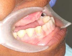
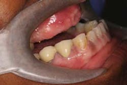
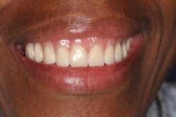
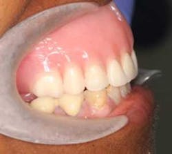
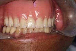
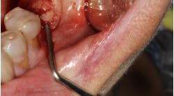
Figures 1 and 2: Patient presents for an implant bridge consultation. It was determined that the patient would need significant bone grafting to repair her horizontal deficiency prior to implant placement. Prosthetically driven implant placement will allow for a restoration that can be screw retained, hygienic, and esthetic.
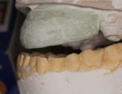
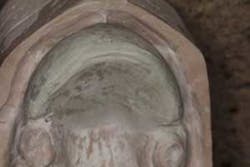
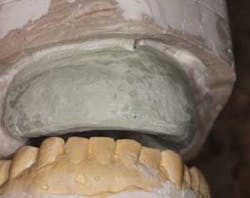
Figures 3 and 4: Models were taken and then modified to create ideal ridge form in stone. This model will facilitate guide preparation for the periodontist to augment the anterior ridge with both hard and soft tissue.
Figure 5:A denture was fabricated based on the altered stone model. This denture will serve several purposes. First, this will give our patient a preview of what the final result can look like after implants and graft integrate. Second, this serves as a guide for the periodontist to determine how much grafting is necessary and where to place the implants. Lastly, the denture will serve as an immediate denture that can be used after ridge augmentation.
Figures 6, 7, and 8:The denture was fabricated and delivered. The patient approved of the esthetics.
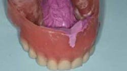
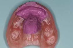
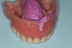
Figures 9 and 10:Polyvinyl siloxane impression material was placed inside the denture to show how much augmentation will be necessary to achieve the proper outcome. The patient was then referred for grafting in the areas indicated in purple.
ADDITIONAL READING |Importance of soft tissue in interdisciplinary cases: A case study involving orthodontics, periodontics, dental implants
