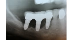
An alternative to conventional dental implants: short implants
Osseointegrated implants have become a routine solution for treating patients who are either completely edentulous, partially edentulous, or missing a single tooth. Studies have confirmed that dental implants have a favorable long-term prognosis as compared to conventional fixed prosthodontics. (1) Despite having high success rates, limitations to single-tooth implant placement and success have been observed in the posterior regions of the dental arches. (2) In the past, implants placed in the posterior region have been associated with higher rates of failure than implant placed the anterior region. (3) Posterior regions of the dental arches generally have less available bone height, poorer bone quality, while, at the same time, teeth in this region are exposed to greater occlusal loads than the anterior regions of the mouth (4) Poorer bone height and quality have been associated with an increased chance of implant failure. (5) Because of the reduced alveolar bone height and density in the posterior regions of the mouth, which often precedes or accompanies tooth loss, (6) anatomic limitations to implant placement, such as the maxillary sinus and the mandibular nerve, exist. Surgical procedures to compensate for this tissue deficiency, such as sinus and/or ridge augmentation procedures, have proved to be successful in providing sufficient bone quantity and quality for implant placement and prosthetic support, (7) However, increased cost, surgical time, morbidity, and healing time are often associated with these procedures. Alternatives to performing these additional augmentation procedures in cases where patients have atrophic posterior regions have come in the form of zygomatic (8) and shortened implants. Although the use of short implants seems to be an obvious alternative in cases where conventional implants are not an option without additional procedures, short implants have been associated with decreased implant success rates. (9) On the other hand, many authors have shown similar success rates with short implants as compared with conventional length implants and attribute these similar success rates to the improvement in both surgical/restorative technique and implant material. (10)
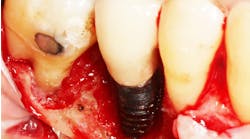
The use of short implants has been proposed as a viable alternative in patients with resorbed posterior regions unwilling to undergo ridge augmentation procedures. In addition to the avoidance of additional surgery, some studies have shown that the benefits of short implants include an easier fixture insertion, a simplified osteotomy preparation, and a decreased potential for overheating the alveolus. (11) These studies also suggest that implant angulation relative to the occlusal load may be improved “since the basal bone beyond the original alveolar ridge for longer implants is not always in the long axis of the missing tooth.” (12) Despite all of the benefits of short implants, the one overwhelming reason why most clinicians are hesitant to use short implants is the fear that reduced implant length may overload the surrounding bone and lead to implant failure. One of the adages of implant dentistry is to “use as many implants as possible of maximum length.” (13) Therefore, the question can be asked: Is there a critical length where implants begin to have increased complications?
According to Bahat, advantages of increased implant length include increased initial stability, long-term resistance to bending moment forces, expedited healing, and a decreased risk of movement at the interface. In one study, Bahat placed 732 implants in the posterior maxilla of partially edentulous patients and reported a failure rate of 9.5% for 7 mm implants as compared with 3.8% for all other lengths. (14) In a series of 13 studies, Goodacre reported on a total of 2,754 implants with a length of 10 mm or less and compared their success rates with 3,015 implants of greater than 10 mm in length. The failure rate of the shorter implant group was 10% compared to 3% in the longer implant group. (15) Naert also reported on clinical outcomes of placing short dental implants and found implants shorter than 10 mm had a survival rate average of 81.5% while implants longer than 10 mm had a 94.1% success rate. (16)
Another study conducted by Ivanoff found that 5 mm x 8 mm implants failed 25% of the time in the maxilla and 33% of the time in the mandible compared with both 5 x10 mm and 5 x 12 mm implants which failed 10% of the time in the maxilla and no reported failures in the mandible. (17) Similarly, Winkler et al published a multicenter report with data collected from 30 hospitals and two university sites during a three-year period. He found that 8, 10, and 13 mm implants had a respective failure rate of 13%, 10.9%, and 5.7%. This study shows that failure rate was directly related to implant length and increased failure rates of 2- to 5-fold can be evident with shorter implants. (18) Another multicenter study conducted by Weng et al showed that 60% of all failed implants were 10 mm or less in length. The 7 mm implant failed 26% of the time, the 8 mm implant had a 19% failure, while the 10 mm implant had a 9% failure. The overall failure rate of all implants in the study was 9%. Therefore, the 10 mm implant survival rate was similar to the longer length implants, while implants shorter than 10 mm demonstrated significantly greater risks of failure. (19) The overall implications of these studies are surprising since shorter implants (defined as less than 1 0mm from these studies) show failure rates as high as 16% to 33% vs. 4% to 9% with conventional length implants. Of importance to note is that the failures in most of these studies were after loading, not failure of the implant to integrate.
On the other hand, there are many studies that support the use of short implants and show that short implants have success rates similar to conventional length implants. Fugazzotto et al obtained an overall cumulative success rate of 95.1% with implants of 7 mm to 9 mm in length in the posterior maxilla. (20) Deporter et al found a 100% success rate for short implants (mean length of 6.9 mm) in the posterior maxilla. (21) Renouard and Nissand placed 96 short implants ranging 6 mm to 8.5 mm in length in the posterior maxilla and found a 94.6% success rate two years after loading. (22) Misch et al placed 437 implants less than 10 mm in length (408 implants were 9 mm long and 29 implants were 7 mm long). The majority of these restorations were in the posterior mandible or maxilla. The restorations in this study were loaded for at least 18 months. Of the 437 implants, there were three implant failures in the posterior mandible and one failure in the posterior maxilla (99% survival). All these failures were implants 9 mm long and 4 mm in diameter. (23) No implants failed during the prosthesis fabrication. Hence, the overall implant survival from Stage 1 surgery to prosthesis delivery was 99.0%. The implants and restorations were followed at least 18 months and up to three years. No implants were lost during this time frame, and no restorations were remade. Misch and Fugazzotto suggest in their articles that unlike previous articles that show a decreased success rate with short implants, their success rates, which are comparable to conventional length implants, are a result of improved surgical technique and implant design.
In summary, the posterior regions of the mouth often have less available bone height than the anterior regions and have traditionally been considered areas where implant success is decreased. The bone density of the remaining bone after tooth loss is often less in the posterior regions than the anterior region of the mouth. Many of the studies reviewed in this paper show implants shorter than 10 mm often have a higher failure rate than longer implants. These complications may be related to an increase in crown height, higher bite forces in the posterior regions, and less bone density. As a result, biomechanical methods to decrease stresses to the implant-bone interface are warranted. The forces to the implants may be reduced by eliminating contacts in lateral excursions, eliminating cantilevers on the prosthesis, increasing thread pitch and depth, and roughening the implant surface. The decrease in stress forces to the prosthesis may be decreased by increasing the implant number, increasing the implant diameter, altering implant design to increase surface area, and splinting the implants together. These alterations in both implant technique and design can account for the similar success rates achieved in many studies between short and conventional length implants.
References
1. Cochran et al. The scientific basis for clinical experiences with Straumann implants including the ITI dental implant system: A consensus report. COIR 2000;11 (supplemental):33-58.
2. Zarb G, Schmitt A. The longitudinal clinical effectiveness of osseointegrated dental implants in the posterior partially edentulous patients. IJPRD 1993;6:189-196.
3. Kopp CD. Branemark osseointegration. Prognosis and treatment rationale. DCNA 1989;33:701-731.
4 Scurria et al. Prognostic variables associated with implant failure: A retrospective effectiveness study. IJOMI 1998;13:400-406.
5. Jaffin RA, Berman CL. The excessive loss of Branemark fixtures in type IV bone. J Perio 1991;62:2-4.
6. Schropp et al. Bone healing and soft tissue contour changes after single-tooth extraction: a clinical and radiographic 12-month prospective study. IJPRD 2003;23 (4):313-323.
7. Wallace SS, Froum SJ. Effect of maxillary sinus augmentation on the survival of endosseous dental implants. A systematic review. Ann Perio 2003;8:328-343.
8. Will not be discussed in this paper.
9. Moy PK, Bain CA. Relation between fixture length and implant failure. JDR 1992;71:637-641.
10. Deporter et al. Simplifying management of the posterior maxilla using short, porous-surfaced dental implants. IJPRD 2000;20:476-485.
11. Misch CE, Bidez MW. Implant protected occlusion, a biomechanical rationale. Compendium. 1994;15:1330,1332,1334.
12. ten Bruggenkate CM, Asikainen P, Foitzik C, et al. Short (6-mm) nonsubmerged dental implants: results of a Multicenter clinical trail of 1 to 7 years. IJOMI. 1998;13:791-798.
13. Balshi et al. A comparative study of one implant versus two replacing a single molar. IJOMI 1996;11:372-378.
14. Bahat O. Treatment planning and placement of implants in the posterior maxilla: Report of 732 consecutive Nobelpharma implants. IJOMI 1993:8:151-161.
15. Goodacre CJ, Bernal G, Rungcharassaeng K, Kan J. Clinical complications with implants and implant prostheses. JPD 2003;9:121-132.
16. Naert I, Quirynen M, van Steenberghe D, Darius P. A six-year prosthodontic study of 509 consecutively inserted implants for the treatment of partial edentulism. JPD 1992;67:236-245.
17. Ivanoff CJ, Grondahl K, Sennerby L, et al. Influence of variations in implant diameters: a 3- to 5-year retrospective clinical report. IJOMI. 1999;14:173-180.
18. Winkler S, Morris HF, Ochi S. Implant survival to 36 months as related to length and diameter. Ann Perio 2000;5:22-31.
19. Weng D, Jacobson Z, Tarnow D, et al. A prospective multicenter clinical trial of 3i machined-surface implants: results after 6 years of follow-up. IJOMI 2003;18:417-423.
20. Fugazzotto et al. Success and failure rates of 9.mm or shorter implants in the replacement of missing maxillary molars when restored with individual crowns: preliminary results 0-84months in function. A retrospective study. J Perio 2004;75:327-332.
21. Deporter et al. Simplifying management of the posterior maxilla using short, porous-surfaced dental implants and simultaneous indirect sinus elevation. IJPRD 2000;20:P476-485.
22. Renouard F, Nisand D. Short implants in the severely resorbed maxilla: A 2 -year retrospective clinical study. CIDRR 2005;7(Suppl):104-110.
23. Misch CE, Bidez MW. Occlusal considerations for implant-supported prosthesis: implant protected occlusion. In: Misch CE. Dental Implant Prosthetics. St. Louis, Mo: Elsevier/Mosby; 2005:472-510.

