Clinical dental advantages of the apically positioned flap
The apically positioned flap is a commonly used surgical approach to achieve pocket elimination. This technique is important for maintaining an adequate zone of keratinized tissue, as opposed to the gingivectomy technique, where soft tissue is resected.
ADDITIONAL READING |Attached gingiva and dental implants
Indications for the apically positioned flap include 1. surgical crown lengthening procedures, and 2. the treatment of periodontal disease where preserving the maximum amount of keratinized gingiva is the desired outcome. The contraindications for the apically positioned flap include root hypersensitivity, high rate of root caries, esthetic limitations, anatomic limitations, inadequate clinical attachment, and deep infrabony defects.
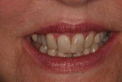
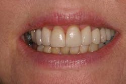
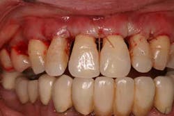
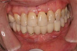
1. Clinical crown lengthening is a surgical procedure to increase the amount of exposed tooth structure. There are two main types of crown lengthening: functional and esthetic. Functional crown lengthening may be performed to expose subgingival caries, a fracture, or both. Garguillo et al. described a concept known as “biologic width,” which consisted of a connective tissue attachment of 1.07 mm and the average epithelial attachment was 0.97 mm, forming an average biologic width of 2.04 mm. Rosenberg et al. furthered this concept and combined the average biologic width of 2.04 mm with the 1 mm to 2 mm essential for restorative dentistry, concluding that 3.5 mm to 4 mm of sound supraosseous crest tooth structure is needed to fulfill the total requirement for restorative dentistry. The indications for crown lengthening surgery include caries, fracture, altered passive eruption, root surface preparations, and restorative requirements. (Figs. 1 and 2) The apically positioned flap offers several advantages over the gingivectomy technique for surgical crown-lengthening procedures. These include maximizing the postoperative keratinized attached tissue, increased access to the alveolar bone, faster and more comfortable wound healing, and more predictable placement of the margin of gingiva.Fig. 1: Clinical image prior to crown lengthening for preparation for restoration
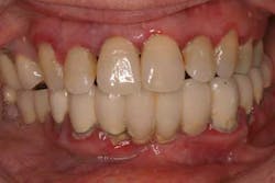
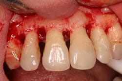
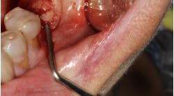
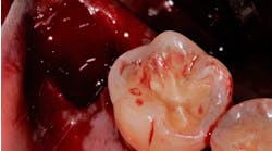
Fig. 2: Post crown lengthening image after temporization 2. When treating periodontal disease, (Fig. 3) the apically repositioned flap technique involves a reverse beveled incision with a scalpel. The distance from the tooth, buccally or lingually, is dependent on the pocket depth, and the beveled incision should be scalloped to maximize the amount of interproximal alveolar bone that will be covered after the flap is positioned apically. The original technique also involves vertical releases at the mesial and distal extent of the flap, extending beyond the mucco-gingival junction, to facilitate the apical positioning of the flap. Next, a full thickness mucco-periosteal flap, including the alveolar mucosa and the keratinized tissue, is raised both buccally and lingually beyond the mucco-gingival junction. (Fig. 4) If the flap is not raised beyond the mucco-gingival junction, then it would be impossible to advance the flap in an apical direction. After the full thickness mucco-periosteal flap is elevated, then the collar of soft tissue is removed from around the necks of the teeth with surgical curettes. This marginal collar of soft tissue includes diseased epithelial soft tissue and granulation tissue. After the surgical area is degranulated, the roots of the teeth are meticulously scaled and root planed.
Fig. 3: Clinical image of a patient with severe periodontal disease
Fig. 4: Mucoperiosteal flap beyond mucogingival junction and surgical debridement
Once the physiologic alveolar bone contour is achieved, the flap is apically advanced to the alveolar bone crest and secured with sutures. (Fig. 5) Since it is not always possible to completely cover the entire alveolar bone complex with soft tissue, some areas of alveolar bone may be left denuded. After the surgical site has healed, the technique should preserve the zone of keratinized soft tissue and yield shallow probing depths. (Fig 6)
Fig. 5: Apically repositioned flap and suture
Fig. 6: Gingival tissues and bone levels remain stable 2½ years after surgical therapy
Photos courtesy of Scott Froum, DDS
References
1. Ainamo A, Bergenholtz A, Hugoson A, Ainamo J. Location of the mucogingival junction 18 years after apically repositioned fl ap surgery. Journal of Clinical Periodontology 1992;19:49-52.
2. Bernimoulin JP, Lüscher B, Muhlemann HR. Coronally repositioned periodontal flap. J Clin Periodontol 1975;2:1-13.
3. Carnio J, Miller P. Increasing the amount of attached gingiva using a modified apically repositioned flap, J Periodontol 1999;70:1110-1117.
4. Donnenfeld OW, Marks RM, Glickman I. The apically repositioned flap — a clinical study. Journal of Periodontology 1964;35:381-387.
5. Friedman N. Periodontal osseous surgery: osteoplastyand ostectomy. Journal of Periodontology 1955;26:257-269.
6. Friedman N. Mucogingival surgery. The apically repositioned flap. Journal of Periodontology 1962;33:328-340.
7. Gargiulo AW, Wentz FM, Orban B. Dimensions and relations of the dento-gingival junction in humans. J Periodontol 1961;32:261-267.
8. Harris, R.J. Clinical evaluation of three techniques to augment keratinized tissue without root coverage. Journal of Periodontology 2001;72:932-938.
9. Herrero F, Scott JB, Maropis PS, Yukna RA. Clinical comparison of desired vs. actual amount of surgical crown lengthening. Journal of Periodontology 1995;66:568-571.
10. Levy RM, Giannobile WV, Feres M et al: The effect of apically repositioned flap surgery on clinical parameters and the composition of the subgingival microbiota: 12-month data. Int J Periodontics Restorative Dent 2002;22:209-219.
11. Machtei E, Ben-Yehouda A. The effect of postsurgical flap placement on probing depth and attachment level: a two-year longitudinal study. J Periodontol 1994;65:855.
12. Nabers CL. Repositioning the attached gingiva. Journal of Periodontology 1954;25:8-39.
13. Pippin DJ. Fate of pocket epithelium in an apically positioned flap. J ClinPeriodontol 1990;17:385-391.
14. Polson AM, Heijl L. Osseous repair in infrabony periodontal defects. Journal of Clinical Periodontology 1978;5:13-23.
15. Pontoriero R, Carnevale G. Surgical crown lengthening: A 12-month clinical wound healing study. Journal of Periodontology 2001;72:841-848.
16. Rosenberg ES, Garber DA, Evian CI. Tooth lengthening procedures. Compend Contin Educ Dent 1980;1:161.
17. Schluger S. Osseous resection — a basic principle in periodontal surgery? Oral Surgery, Oral Medicine and Oral Pathology 1949;2:316-325.
18. Townsend-Olsen C, Ammons W, van Belle G. A longitudinal study comparing apically repositioned flaps, with and without osseous surgery. Int J Periodontics Restorative Dent 1985;4:11-34.
19. Vanarsdall R, Corn H. Soft tissue management of labially positioned unerupted teeth. Am J Orthod 1977;72:53-64; Oral Pathology 2:316–325.
20. Vermette M, Kokich V. Uncovering labially impacted teeth: apically positioned flap and closed-eruption techniques. Angle Orthod 1995;65:23-34.

