A review of available periodontal surgical techniques for pocket elimination
Since the mid-1900s we’ve known that soft-tissue resection alone fails to eliminate periodontal pockets. Drs. James Ramos and Scott Froum give an overview of the current surgical techniques for periodontal pocket elimination: curettage, gingivectomy, osseous surgery through the palatal flap, and laser-assisted pocket-reduction treatment.
PERIODONTAL SURGERY was first described in 1924 and again in 1935, but it did not gain acceptance until 1949 when Saul Schluger, DDS, set forth his principles of harmonious gingival and osseous contour for the maintenance of pocket elimination. (1) Schluger stated that although conservative treatment is feasible and preferable in comparison to surgery, this conservative approach might lead to tissue breakdown because soft-tissue resection alone fails to eliminate pockets. This article will review surgical techniques available for periodontal pocket elimination.
Surgical techniques for periodontal pocket elimination
Curettage
Barkann et al. (2) described the procedure known as curettage, the surgical elimination of the epithelial lining of the pocket. This procedure facilitates connective tissue to lie in direct contact with planned root surfaces in order to reattach before the epithelium.
Gingivectomy
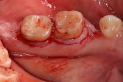
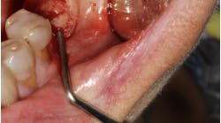
The gingivectomy (figure 1) is a technique used to excise excess gingiva without much consideration to shaping the underlying bone. This tissue is removed to the base of the underlying bone, and the new tissue is allowed to granulate in. Minor adjustments can be made to the bone but usually without flap reflection. (3)
Figure 1: Gingivectomy
Osseous surgery: palatal approach
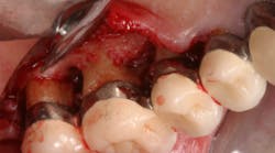
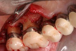
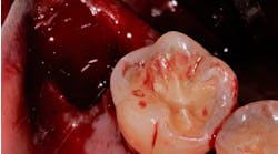
First described by Ochsenbein et al., osseous surgery through the palatal approach (figure 2) utilizes the traditional principles of osseous surgery (full-thickness flap with bone osteoplasty) in order to create positive architecture, but access is through the palatal flap leaving the buccal tissue intact. (4) This approach allows for better esthetics, elimination of potential surgically created recession defects, and better embrasure form in the palatal aspect. In addition, the buccal roots of the maxillary molars can be prominent and fenestrated in the alveolar housing. Surgical exposure of an already thin cortical plate may result in heightened bone loss and a greater soft-tissue problem. (5) Careful attention should be paid to the amount of osteoplasty actually necessary, since by just raising the flap alone, bone loss of about 0.8–1 mm is to be expected. (6)
Figure 2: Osseous surgery through the palatal approach
Laser-assisted pocket-reduction treatment
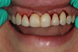
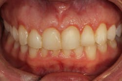
Many different types of lasers are used for pocket-reduction treatment in periodontal therapy (figures 3a–3c). Nd:YAG lasers, Er:YAG lasers, Er,Cr:YSSG lasers, 10.6-micron carbon dioxide lasers, 9.3-micron carbon dioxide lasers, and diode lasers all have been used to reduce periodontal pockets. Most lasers have soft-tissue removal indications, while other lasers—such as those in the Er:YAG family and the 9.3-micron carbon dioxide lasers—have capabilities of both hard- and soft-tissue removal. Lasers are a useful adjunct to the traditional oral surgery armamentarium and have great success in case-specific, operator-familiar circumstances. The best laser is always the one that works the best for the particular clinician and patient case.
Figure 3a: Inflamed gingiva leading to pseudo pocket
Figure 3b: Pocket reduction with 10.6-micron CO2 laser
Figure 3c: Three-month follow-up
Conclusion
In conclusion, many different surgical techniques have been described in the literature that have the ability to eliminate periodontal pockets. Patient compliance, practitioner experience, and case selection continue to be important factors in determining which surgical technique will work best.
References
1. Schluger S. Osseous resection—A basic principle in periodontal surgery. Read for the Section on Periodontia at the American Dental Association. Chicago, IL. September 1948. Oral Surg, Oral Med, Oral Pathol. 1949;2(3):316-325. doi:10.1016/0030-4220(49)90363-1.
2. Barkann L. Conservative surgical technique for the eradication of a pyorrhea pocket. J Am Dent Assoc. 1939;26(1):61-68.
3. Beube FE. Interdental tissue resection: An experimental study of a surgical technique which aids in repair of the periodontal tissues to their original contour and function. Oral Surg, Oral Med, Oral Pathol. 1947;33:497-504.
4. Ochsenbein C, Bohannan HM. The palatal approach to osseous surgery. J Periodontol. 1963;34(1):60-68.
5. Goldman HM, Cohen DW. Periodontia. 4th ed. St. Louis, MO: C. V. Mosby Co.; 1957:195.
6. Donnenfeld OW, Hoag PM, Weissman DP. A clinical study on the effects of osteoplasty. J Periodontol. 1970;41(3):131-141. doi:10.1902/jop.1970.41.3.131.
Scott Froum, DDS, a graduate of the State University of New York, Stony Brook School of Dental Medicine, is a periodontist in private practice at 1110 2nd Avenue, Suite 305, New York City, New York. He is the editorial director of Perio-Implant Advisory and serves on the editorial advisory board of Dental Economics. Dr. Froum, a diplomate of the American Board of Periodontology, is a clinical associate professor at SUNY Stony Brook School of Dental Medicine in the Department of Periodontology. He serves on the board of editorial consultants for the Academy of Osseointegration's Academy News. Contact him through his website at drscottfroum.comor (212) 751-8530.
