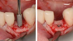
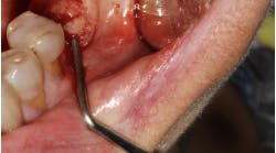
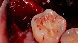
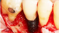
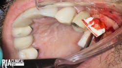
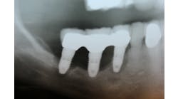
Narrow implants (from 3.5 mm in diameter and less) have been designed for treatment of the anterior maxillary and mandibular regions mainly for the replacement of lower incisors, but not for the replacement of premolars and molars. Concerning these teeth, wider implants are indicated.1
At the beginning of the 1990s, due to their mechanical properties, 5 mm diameter implants were considered the ideal implants for the replacement of premolars. A study published in the late 1990s disproved this belief, reporting higher failure rates (18%) for 5 mm diameter implants compared to 3.75 mm and 4 mm wide implants, which yielded 5% and 3% failure rates respectively.2 Similarly, other authors observed a higher survival rate using 3.75 mm diameter implants rather than adopting wider diameter ones.3
Moreover, long-term results show more marginal bone loss around wide-diameter implants than around 3.75 mm or 4 mm diameter implants.4
Then, one should ask the following question: do we need more bone and less titanium, or less bone and more titanium?
Some authors concluded that narrow implants (2.75 mm to 3.25 mm diameter) could be successfully used as a minimally invasive alternative to horizontal bone augmentation in posterior mandibles for up to one year of function.5
The logical evidences in such cases would be more bone to promote osseointegration.
In 1998, I started to place narrow implants not only in the anterior areas, but also in posterior maxillary and mandibular regions in order to replace premolars and molars. I began to treat partially edentulous patients with a high success rate.
• simplification of the surgical procedures
• reduced grafting procedures
• reduced morbidity and first complications
• reduced postsurgical problems
I immediately found an increasing number of cases to be treated in this way. From 1998 to 2008, I placed 824 narrow implants, with a success rate of 98.5%. From 2009 to 2016, I placed 4,266 narrow implants, multiplying the number of narrow implants by nearly six.
During these years, in my clinical experience, I found:
• a high success rate
• a very stable marginal bone level
• one implant fracture
Placement of narrow implants. In a full-arch restoration in the presence of alveolae and thin ridges, narrow implants allow the placement of more biomaterial in the alveolae for ridge preservation. Bone grafting will be avoided in the presence of the ridges.
Literature review
When narrow implants were placed in both posterior jaws and the values for marginal bone resorption at one, five, and 10 years were measured, they were within the accepted standard success criteria for dental implants. Regarding the implant failures, the majority occurred in the first six months of function, following the pattern for standard-diameter implants.6
A recent systematic review reports average survival rates of mini-implants (1.8 mm to 2.9 mm diameter) and narrow implants (3 mm to 3.5 mm diameter) as 98% and 98% respectively, while the average success rates were 93% and 96% respectively. The average peri-implant bone loss after 12, 24, and 36 months was 0.89 mm, 1.18 mm, and 1.02 mm for mini-implants and 0.18 mm, 0.12 mm, and -0.32 mm for narrow implants. Both mini-implants and narrow implants showed adequate clinical behavior as overdenture retainers and clinical outcomes similar to standard implants. The narrow implants also showed a higher survival rate for the studies with higher follow-up time, and they present a better long-term predictability than the mini-implants when conventional loading is applied.4
Another review concludes that survival rates reported for narrow implants are analogous to those reported for standard-width implants. These survival rates did not appear to differ between studies that used flapless and flap-reflection techniques. The failure rate appeared to be higher in shorter than in longer ones in the studies in which the length of the failed implants was reported. Narrow implants could be considered for use with fixed restorations and mandibular overdentures, since their success rate appears to be comparable to that of regular-diameter implants.7
Some authors state that using small-diameter implants is a treatment option as predictable as using standard-diameter implants. They summarize their findings as follows:8
• The medium-term prognosis of narrow implants is comparable to that of standard-diameter implants followed up in the present study. Therefore, the high reliability of small-diameter implants is confirmed.
• Standard- and narrow-implant prognoses were influenced by peri-implant bone infection more than biomechanical factors, such as implant overloading.
• Peri-implant bone resorption was not significantly influenced by different implant diameters (3.3 mm and 4.1 mm).
• Bone quality seems to be an important prognosis factor both for standard- and small-diameter implants; spongy bone (type 4) may increase implant failures.
• Survival of standard and narrow implants does not seem to be affected by implant location.
Conclusion
Narrow implants can be used in almost every area, except the posterior maxilla/mandibula with a limited bone height. The use of narrow implants is a safe and reliable technique with no problems in relation to function and esthetics.
References
- Davarpanah M, Martinez H, Tecucianu JF, Celletti R, Lazzara R. Small-diameter implants: indications and contraindications. J Esthet Dent. 2000;12(4):186-194.
- Ivanoff CJ, Gröndahl K, Sennerby L, Bergström C, Lekholm U. Influence of variations in implant diameters: a 3- to 5-year retrospective clinical report. Int J Oral Maxillofac Implants. 1999;14(2):173-180.
- Shin SW, Bryant SR, Zarb GA. A retrospective study on the treatment outcome of wide-bodied implants. Int J Prosthodont. 2004;17(1):52-58.
- Marcello-Machado RM, Faot F, Schuster AJ, Nascimento GG, Del Bel Cury AA. Mini-implants and narrow diameter implants as mandibular overdenture retainers: A systematic review and meta-analysis of clinical and radiographic outcomes. J Oral Rehabil. 2018;45(2):161-183. doi:10.1111/joor.12585
- Grandi T, Svezia L, Grandi G. Narrow implants (2.75 and 3.25 mm diameter) supporting a fixed splinted prostheses in posterior regions of mandible: one-year results from a prospective cohort study. Int J Implant Dent. 2017;3(1):43. doi:10.1186/s40729-017-0102-6
- Maló P, de Araújo Nobre M. Implants (3.3 mm diameter) for the rehabilitation of edentulous posterior regions: a retrospective clinical study with up to 11 years of follow-up. Clin Implant Dent Relat Res. 2011;13(2):95-103. doi:10.1111/j.1708-8208.2009.00188.x
- Sohrabi K, Mushantat A, Esfandiari S, Feine J. How successful are small-diameter implants? A literature review. Clin Oral Implants Res. 2012;23(5):515-525. doi:10.1111/j.1600-0501.2011.02410.x
- Romeo E, Lops D, Amorfini L, Chiapasco M, Ghisolfi M, Vogel G. Clinical and radiographic evaluation of small-diameter (3.3-mm) implants followed for 1-7 years: a longitudinal study. Clin Oral Implants Res. 2006;17(2):139-148. doi:10.1111/j.1600-0501.2005.01191.x
Patrick Palacci, DDS, obtained his DDS degree in 1975 at Aix-Marseille University, in Marseille, France. He continued his postgraduate education in periodontology at Boston University from 1977 to 1981. In 1982, he was appointed as a visiting professor at Boston University. Dr. Palacci is a private practitioner and the head of the Brånemark Osseointegration Center in Marseille. He is the author of numerous scientific articles and textbooks related to esthetic and implant dentistry and soft-tissue management. He now conducts a number of courses in his private clinic and is an invited speaker to scientific meetings around the world.
